Development of novel applications of x-ray computed tomography in plant science

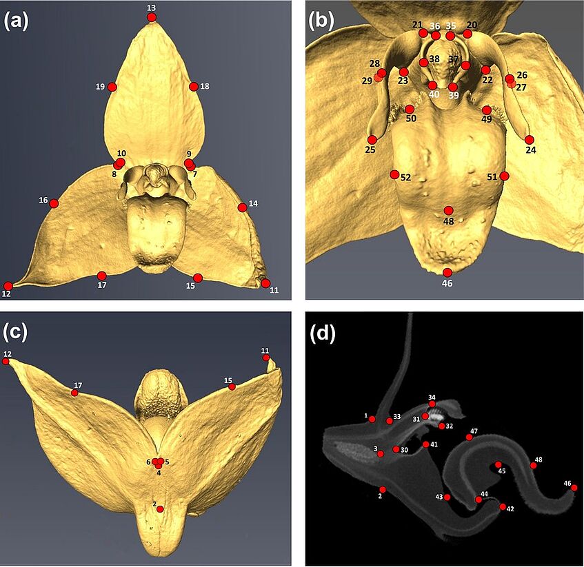
Landmarks (LMs 1–52; red dots) placed onto high-resolution X-ray computed tomography (HRX-CT) scans of an exemplary flower of Malagasy Bulbophyllum (B. francoisii of sect. Elasmotopus) (© Artuso et al. 2022, Fig. 2)
High resolution x-ray computed tomography remains strongly underused in plant sciences despite of its high potential in delivering detailed 3D phenotypical information.
Our lab has recently developed efficient protocols to study plant material via tomography and we have applied these protocols in a series of innovative, collaborative studies in the fields of comparative floral morphology, floral development, pollination biology, and plant interactions, and paleobotany.

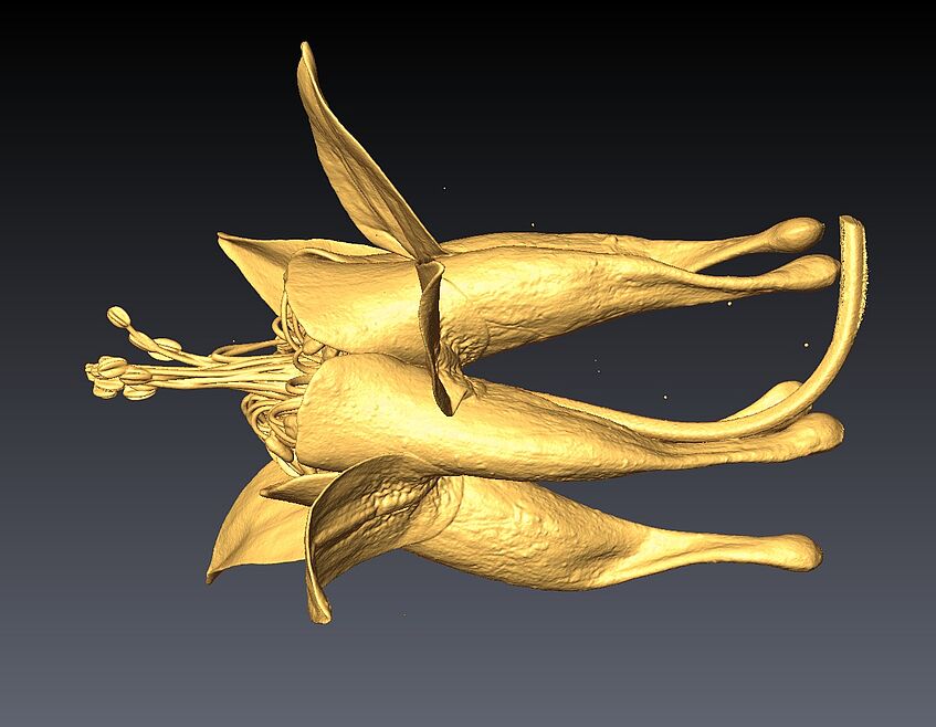
Aquilegia canadensis, anthetic flower observed by micro computer tomography © M. Chartier

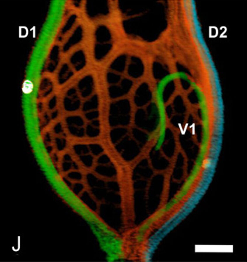
Ovary with ovule (HRXCT, 2D reconstructions in longitudinal sections) of the pistillate flower of Celtis species, C. occidentalis (© Leme et al. 2021, Fig. 8J)
